
5 signs of a base of skull fracture
A base of skull fracture is a break in one or more bones of the skull floor (yes, that’s bones plural; there’s 22!). Trauma doctors care about recognising these because they indicate significant head trauma, and potential brain injury. Such patients are often unable to give a good history, through reduced consciousness.
These types of fractures can give 5 good clinical signs at a glance, that are useful by suggesting significant trauma. The signs are summarised above, and broken down below:
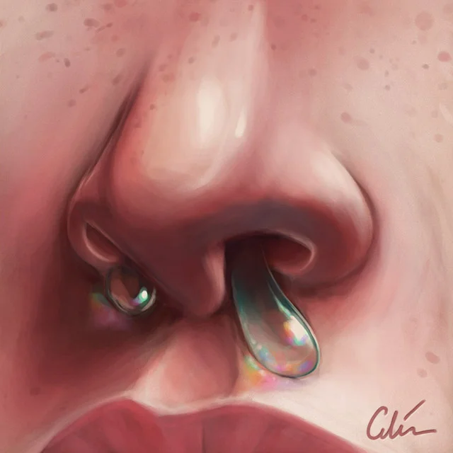
CSF rhinorrhoea / otorrhoea:
Cerebrospinal fluid (CSF) is the clear liquid that the brain is submerged in, and cushioned by in the skull. The meninges are waterproof fibrous layers between the inside of the skull and this fluid, keeping it from leaking through. A crack in the skull can tear the meninges, resulting in a leak of CSF.
Depending on where this leak happens, CSF can be seen coming from the nose or ear, which are two nearby exit routes. Blood may also drip out, but it’s less specific than this clear fluid in suggesting a base of skull fracture.
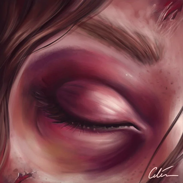
Raccoon eyes (periorbital ecchymoses):
When a base of skull fracture involves the orbit (the eye socket bones), then bleeding can occur from these, into the soft tissues around the eyeballs. This is seen as bruising around the eyes bilaterally (both sides involved). This can take a few days to develop, unlike ‘black eyes’ which tend to appear within hours.
Other differences include ‘tarsal plate sparing’ (which tends to occur in raccoon eyes), an area of the upper eyelid without bruising. This is due to the fibrous tarsal plate (which helps prevent the eyelid inverting when blinking), which impedes the spread of bruising. Raccoon eyes bruising tends to be confined to the orbit, whereas a black eye may spread outside this area.
Haemotympanum: Cracks in the base of the skull can involve the inner ear canal. Bleeding here can result in trapped blood behind the ear drum, which can be seen with a handheld Otoscope (oto means ear in latin). The pressure of this pushes out the normal concave resting shape of the ear drum to be convex.
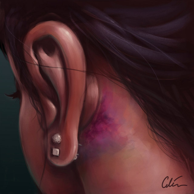
Battle sign: This is bruising seen behind the ear, over a part of the skull called the ‘mastoid process’, where the big sternocleidomastoid muscle of the neck inserts. Direct trauma to this area could of course cause this too, but you might expect skin evidence such as a laceration, or clear history that this was the case.
This illustration is a reminder of the real injury these signs are clues to: internal head trauma. The inside of the skull has lots of sharp edges to lacerate a brain. High velocity trauma can cause bruising of the brain on the side of the trauma, and also the opposite, called ‘contre-coup’ as the brain is bounced around.
When tissue is damaged it tends to swell. As there isn’t much room for expansion within the skull, pressure in the skull increases rapidly with swelling. This can quickly impede arterial blood flow to the brain, leading to hypoxic injury, strokes, and death.
Well that’s it for my illustrated summary of visual clues hinting a base of skull fracture. A special thanks goes out to Dr Doug Murray (Consultant Emergency Physician and Clinical Toxicologist), who kindly shared his insights to improve the article.
Please share with anyone you think might be interested, and let me know how you found the breakdown.