
Schistosomiasis
This art was for a case monograph about hepatic schistosomiasis, created to show how this can result in portal hypertension.

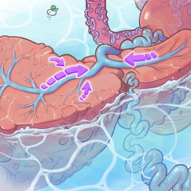
Medical illustration shows the pathogenesis of presinusoidal portal hypertension in schistosomiasis.
Host animals including cattle, dogs, pigs, horses, and goats serve as reservoirs for S japonicum, whereas S mansoni is primarily a human parasite. Schistosoma eggs hatch in freshwater and release miracidia (larvae), which swim and penetrate the snail intermediate host.
Here, sporocysts develop and free-swimming infectious cercariae are released into the water. These penetrate the skin of humans, losing their tail and becoming schistosomula. Schistosomula migrate into the liver portal tracts and mature into adult worms, which then inhabit mesenteric venules of the bowel and rectum.
The mature worms lay eggs; most reach the intestinal lumen, but some are carried to the liver and embed in and obstruct the portal venous system. Untreated or incompletely treated disease can lead to intrahepatic presinusoidal portal hyper-tension, a noncirrhotic cause of portal hypertension. This may be complicated by variceal bleeding, which can be fatal.
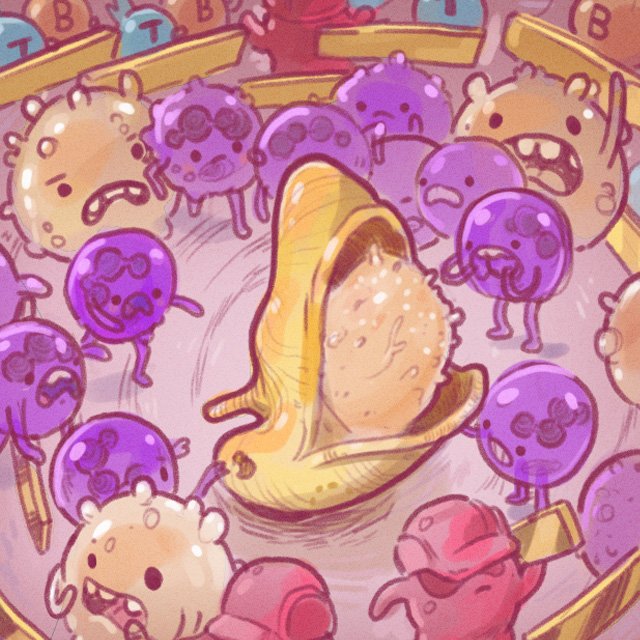
This is the 15th case I’ve illustrated for the ‘Cases from the Cooky Jar’ series in RadioGraphics, and you can read the publication I co-authored with Dr’s Sailer, McDermott, and Huynh here: https://doi.org/10.1148/rg.220193

Read more from the series here